


 Close
Close
 6th July 2026
Venous Thromboembolism
Background
Venous thromboembolism (VTE), occurs when a blood clot forms in a vein. The two main components are deep vein thrombosis and pulmonary embolism. In a deep vein thrombosis (DVT) a thrombus forms in the deep veins of the body, most commonly in the lower limb (legs and pelvis), although it can occur in the upper limbs, cerebral veins, and splanchnic veins as well. This thrombus can break off and embolise, or lodge itself, into a blood vessel downstream. When a thrombus blocks a vessel in the pulmonary arterial tree, this is known as a pulmonary embolism (PE), which can be life-threatening.
Epidemiology
VTE is a very common occurrence worldwide. Within the UK, there is an estimated incidence of 1-2 per 1000 individuals. It can be both the presenting complaint of a patient (patient related) as well as a complication or incidental occurrence whilst in hospital (admission related). As such, it is essential to be able to assess patient risk for VTE and follow the appropriate guidelines for VTE prophylaxis. This is done by weighing the risk of thrombosis against the risk of bleeding. Risk factors for thrombosis can be divided into patient related or admission related.
Patient related risk factors
- Active cancer or treatment for cancer
- Age over 60 years
- Thrombophilia
- Obesity (BMI > 30)
- One or more significant medical comorbidities
- Personal or family history (first degree relative) of VTE
- Medications, including Hormone replacement therapy and contraception containing oestrogen
- Dehydration
- Pregnancy
- Less than 6 weeks postpartum
Admission related risk factors
- Hip or knee replacement
- Hip fracture
- Significantly reduced mobility for 3 days or more
- Total anaesthetic + surgical time > 90 minutes
- Surgery involving the pelvic region or lower limbs with a total anaesthetic + surgical time > 60 minutes
- Acute surgical admission with inflammatory or intra-abdominal pathology
- Critical care admission
- Surgery with significant reduction in mobility
Aetiology
There are 3 major components to take into consideration when thinking about the pathophysiology of VTE. These can be summarised as Virchow’s triad, the components of which include vessel injury, stasis, and hypercoagulability. Any number of procedures, medical conditions, and medications can contribute to these factors and, in turn, lead to thrombosis.
- Vessel injury: Surgery, smoking, hypertension, trauma, catheter (PICC line) insertion
- Stasis: Immobility (may be secondary to surgery), venous insufficiency, varicose veins, obstruction (tumours, pregnancy, obesity)
- Hypercoagulability: Malignancy, pregnancy, inherited thrombophilia, infection/sepsis
Deep vein thrombosis
Diagnosis
Suspicion for DVT is based on history and clinical presentation. Signs and symptoms include pain and tenderness over the affected area. NICE guidelines suggest the following pathway in the initial diagnosis and management of DVT:
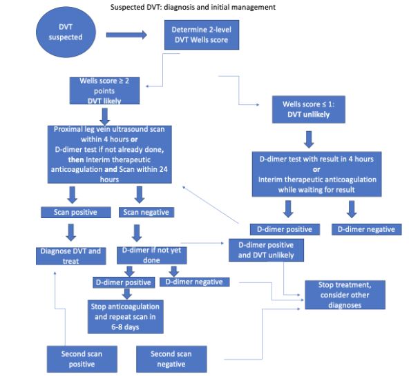
Figure 1: Suspected DVT diagnosis and initial management, adapted from NICE guidelines: Venous thromboembolic diseases: diagnosis, management and thrombophilia testing
Image Source: Wikimedia Commons (2023). Visual Summary, Licensed under a creative commons licence. Available for free at: https://www.nice.org.uk/guidance/ng158/resources/visual-summary-pdf-11193380893 (Accessed 8 May 2025).
| Clinical feature | Points |
|---|---|
| Active cancer (patient either receiving treatment within the last 6 months, or is palliative) | 1 |
| Paralysis, paresis, or (recent) plaster immobilisation of the lower limbs | 1 |
| 1 | |
| Localised tenderness along the distribution of the deep venous system | 1 |
| Swelling of the entire leg | 1 |
| Swelling of the calf at least 3 cm larger than on the asymptomatic side (to be measured 10 cm below the tibial tuberosity) | 1 |
| Pitting oedema of only the symptomatic side | 1 |
| Collateral superficial veins (non-varicose) | 1 |
| History of previous DVT | 1 |
| Alternative diagnosis at least as likely as DVT | -2 |
| DVT likely | 2 points or more |
| DVT unlikely | 1 point or less |
Table 5- 2 Level DVT Wells Score (adapted from: https://www.nejm.org/doi/full/10.1056/NEJMoa023153)
Investigation
Patients will require different investigations depending on which leg they follow on the flow chart. All patients being investigated for a DVT should have baseline blood tests done – full blood count, renal and hepatic function, prothrombin time (PT), and activated partial thromboplastin time (APTT). If starting interim therapeutic anticoagulation, do not wait for blood results. Other mainstay investigations may include: proximal leg vein ultrasound, D-dimer test, CTPA, and V/Q scan. If suspecting a PE, patients will need a chest X-ray and ECG.
Management
Whilst investigating a potential DVT, patients may already have been started on interim therapeutic anticoagulation whilst awaiting further results. If confirmed, this remains the first line treatment for both DVTs and PEs. The standard anticoagulation used is a direct oral anticoagulant (DOAC), or low molecular weight heparin (LMWH) if DOACs are not suitable. There are several contraindications to DOACs. These are summarised below:
- Mechanical heart valves
- Renal impairment* (check local guidelines)
- Triple positive antiphospholipid syndrome
- Severe active bleeding or risk factors for major bleeding
- Pregnancy and breastfeeding
- Liver disease
- Active malignancy
- Allergy/hypersensitivity to drug or its excipients
*A patient with renal impairment is not automatically unsuitable for a DOAC. You need to check the creatinine clearance of the patient which will determine suitability. As per NICE guidelines:
CrCl of 15-50 ml/min can be given:
- Apixaban
- Rivaroxaban
- LMWH fLMor a minimum of 5 days then
- Edoxaban or
- Dabigatran if CrCl ≥ 30 ml/min.
- LMWH or UFH and a Vitamin K antagonist (VKA) for a minimum of 5 days or until INR is at least 2.0 on 2 consecutive measurements, then a VKA alone.
CrCl < 15ml/min can be given:
- LMWH
- UFH
- LMWH or UFH and a VKA for a minimum of 5 days or until INR is at least 2.0 on 2 consecutive measurements, then a VKA alone.
Thrombolytic therapy may be used in symptomatic iliofemoral DVT. Treatment is continued for at least 3 months if DVT/PE is confirmed and may be continued for up to 6 months if the VTE was unprovoked. Blood results will guide further treatment regimens.
In a patient presenting with an unprovoked VTE, patients should have a review of their medical history and baseline bloods, including FBC, renal and hepatic profiles, and a clotting screen. If they do not have any signs or symptoms of cancer, further investigation for any malignancy is not suggested. If there is no clear underlying cause, thrombophilia testing is appropriate. Tests for thrombophilia may be affected by anticoagulation, so specialist advice may be required.
As always, it is important to consider relevant comorbidities, contraindications, and patient preference. Important factors to consider include patient bodyweight, haemodynamic instability, any renal impairment, including renal failure, active cancer, pregnancy/breastfeeding, and patients with triple antiphospholipid syndrome. Results of blood tests will guide further treatment regimens.
If anticoagulation is contraindicated for whatever reason, alternative treatments include mechanical interventions – inferior vena cava (IVC) filters, graduated compression stockings, and percutaneous mechanical thrombectomy. To give a brief explanation of these methods:
- IVC filters are small devices made from metal that are inserted into the IVC. It then traps the clot.
- Graduated compression stockings apply pressure, or, as the name implies, compress your legs to encourage blood to flow back up to the heart, rather than the blood pooling in your legs.
- Percutaneous mechanical thrombectomy is similar to IVC filters in that a device is passed through your veins. Rather than trapping the clot, however, it dissolves the clot into small pieces.
Pulmonary Embolism
As was previously explained, this occurs when a thrombus embolises and lodges into the pulmonary arterial tree. Common signs and symptoms of a PE include dyspnoea, chest pain – often pleuritic, and tachypnoea. In severe cases, patients may have hypotension and loss of consciousness. There may also be overlap with symptoms of a DVT. Investigation, similarly of note – a CTPA is not appropriate for certain patients – patients with allergies to contrast media, severe renal impairment (estimated creatinine clearance less than 30 mls/min), or high risk for irradiation.
Investigation
| Clinical signs and symptoms of DVT (minimum of leg swelling and pain on palpation of the deep veins) | 3 |
|---|---|
| An alternative diagnosis is less likely than PE | 3 |
| Tachycardia (Heart rate more than 100 beats per minute) | 1.5 |
| Immobilisation for more than 3 days OR surgery within the previous 4 weeks | 1.5 |
| Previous history of either DVT or PE | 1.5 |
| Haemoptysis | 1 |
| Malignancy (on treatment, has been on treatment within the last 6 months, or palliative) | 1 |
| PE likely | More than 4 points |
| PE unlikely | 4 points or less |
Table 6- 2 Level PE Wells Score (adapted from: https://bmjopenquality.bmj.com/content/suppl/2015/12/08/bmjquality.u208698.w4222.DC1/ds6617.pdf)
Management
The treatment of a PE is largely the same as DVT – anticoagulation, taking into consideration the same contraindications, comorbidities, and patient preferences as stated previously. Treatment will be continued for at least 3 months. IVC filters and thrombolytic therapy are other candidates for treatment if anticoagulation is contraindicated or not tolerated.
In the event that a patient is haemodynamically unstable, patients should be managed with a continuous unfractionated heparin infusion with a consideration for thrombolytic therapy.
Indications for thrombolytic therapy are:
- Patients with symptomatic iliofemoral DVT with symptoms lasting less than 14 days,
- and good functional status
- and life expectancy of at least 1 year
- and low bleeding risk.
Treatment should be started immediately and not be delayed by imaging or other investigations in the event massive PE is suspected.
VTE in pregnancy and malignancy
Investigation and management of VTE in pregnancy
Pregnancy, in and of itself, is a risk factor for thromboembolism. In preparation for delivery, the body goes into a procoagulant state, to combat the risk of any haemorrhage during or after delivery.
If DVT is suspected, compression duplex ultrasound of the leg should be done. If this is negative AND there is low clinical suspicion, anticoagulation can be stopped.
In all women presenting with signs and symptoms of PE, ECG and CXR should be performed, as well as baseline bloods. If there is concurrent presentation of DVT, duplex ultrasound should be performed. Otherwise, V/Q scan or CTPA should be done. If the CXR is abnormal, CTPA is preferential to a V/Q scan. Both CTPA and V/Q scans will expose the patient and foetus to ionising radiation with V/Q scans causing a slight increase in the risk of cancer developing in childhood but producing a lower dose of radiation to breast tissue (which is more sensitive to radiation during pregnancy). The absolute risk for both is minimal, however patients should be counselled on the risks and consented:
V/Q scans produce a lower dose of radiation to breast tissue (breast tissue is more sensitive to radiation during pregnancy) but carries a slightly increased risk of childhood cancer
CTPA scans will give a smaller dose of radiation to the foetus
The first line management for VTE is low molecular weight heparin (LMWH), and should be started if DVT/PE is clinically suspected, even if imaging has not yet been performed (unless there is a strong contraindication to treatment). Continue to treat with therapeutic doses of subcutaneous LMWH throughout the rest of the pregnancy, and for 6 weeks postnatally – a minimum total of 3 months. In the haemodynamically unstable patient, rapid assessment by the on-call team (including the consultant obstetrician) is crucial. Patients with a massive PE who are haemodynamically unstable should be treated with IV unfractionated heparin.
The same mechanical treatments are available for use in pregnancy under certain circumstances, and would require specialist input.
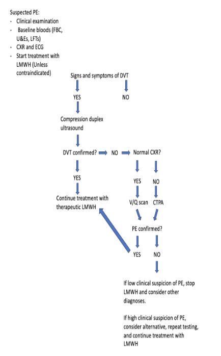
Figure 4 – Algorithm for the investigation and initial management of suspected PE in pregnancy and puerperium. (Adapted from the RCOG guidelines for Thromboembolic Disease in Pregnancy and Puerperium: Acute Management: https://www.rcog.org.uk/media/wj2lpco5/gtg-37b-1.pdf
VTE in Malignancy
Malignancy, like pregnancy, is a risk factor for VTE. Chemotherapy increases the risk sevenfold, compared to patients without cancer. It would be wise to have a low threshold for suspicion of VTE. Investigations should follow the guideline as per above. Management is a complex decision that should be discussed with the MDT. If DVT/PE is confirmed, consider treating with a DOAC. If inappropriate, consider treatment with LMWH +/- a vitamin K antagonist (LMWH is usually discontinued once INR is >2, and warfarin then continued as a monotherapy).
Complications
The commonest complications of a DVT include a PE and post-thrombotic syndrome. In post-thrombotic syndrome, a chronic condition, the thrombus obstructs blood flow, resulting in venous hypertension and valvular incompetence. This can lead to chronic venous insufficiency. As a result, patients may have leg pain, oedema, a feeling of “heaviness,” and skin hyperpigmentation.
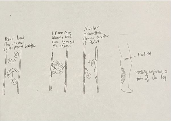
Figure 5 – Pathophysiology of post-thrombotic syndrome
Complications of a PE can be life threatening, and include recurrent thromboembolisms, right heart failure, and cardiogenic shock. When a thrombus occludes a pulmonary artery, it may cause a part of the lung to infarct. This is rare as the lung’s bronchial circulation is not disrupted and is generally adequate. However, if there is an infarct, the pressure in the pulmonary arterial bed may rise, causing cardiac output to drop. Eventually, the patient may go into shock.
Complications related to treatment
- Bleeding
- Heparin induced thrombocytopaenia
Important considerations for VTE treatment (PE and DVT)
PE with haemodynamic instability
- Unfractionated heparin infusion or thrombolysis
Renal impairment, active malignancy, or antiphospholipid syndrome
Medications may be unlicenced so seek specialist advice
Renal impairment: calculate creatinine clearance and prescribe as per NICE or local trust policy
Active cancer: consider initial treatment with DOAC
Antiphospholipid syndrome: LMWH and a vitamin K antagonist (e.g., warfarin) for either 5 days or until INR > 2 on two consecutive readings, then vitamin K antagonist alone
Body weight
- If patient is <50 kg or >120kg, consider monitoring of therapeutic levels of anticoagulation – seek specialist advice
Investigations to guide treatment
- Full blood count, renal function, liver function, coagulation screen (PT, APTT, INR)
- Do not delay urgent treatment whilst awaiting results, but may need doses adjusting subsequently
Medical management for patients without renal impairment, active malignancy, antiphospholipid syndrome, who are haemodynamically stable
- Apixaban and rivaroxaban advised by NICE
- If contraindicated, refer to trust guidelines and seek specialist advice
Length of treatment
- At least 3 months of treatment should be considered for the majority of patients
- Patients with underlying comorbidities such as active malignancy may require longer duration of treatment (up to 6 months). Treatment should be reviewed after 3 months
Monitoring
- Requires monitoring by a community or hospital team, depending on local services and policy
- Self-monitoring of INR and dose adjustment should not be routinely offered
Treatment failure
- If treatment fails (i.e., patient develops a further VTE on treatment), check compliance with treatment, screen for other causes of hypercoagulability, consider dose adjustments, or consider changing to an alternative anticoagulant
References
- B. Paul Herring, Lowen, D., Ho, P. and Hodgson, R. (2023). A systematic review of venous thromboembolism mechanical prophylaxis devices during surgery. Langenbeck’s Archives of Surgery, 408(1). doi:https HYPERLINK “https://doi.org/10.1007/s00423-023-03142-6”://doi.org/10.1007/s00423-023-03142-6.
- Bao, Y., Wan, X., Fu, J. and Wu, B. (2021). The risk of venous thromboembolism in cancer patients receiving chemotherapy: a meta-analysis with systematic review. Annals of Translational Medicine, 9(4), pp.277–277. doi:https://doi.org/10.21037/atm-20-3292.
- British Medical Journal (2022). Pulmonary embolism – Symptoms, diagnosis and treatment | BMJ Best Practice. [online] bestpractice.bmj.com. Available at: https://bestpractice.bmj.com/topics/en-gb/3000115?q=Pulmonary%20embolism&c=suggested
- Farrell, J.J., Sutter, C., Tavri, S. and Patel, I. (2016). Incidence and interventions for post-thrombotic syndrome. Cardiovascular Diagnosis and Therapy, 6(6), pp.623–631. doi:https://doi.org/10.21037/cdt.2016.11.22.
- NICE (2020). Recommendations | Venous thromboembolic diseases: diagnosis, management and thrombophilia testing | Guidance | NICE. [online] www.nice.org.uk. Available at: https://www.nice.org.uk/guidance/ng158/chapter/Recommendations#diagnosis-and-initial-management.
- Nicholson, M., Chan, N., Bhagirath, V. and Ginsberg, J. (2020). Prevention of Venous Thromboembolism in 2020 and Beyond. Journal of Clinical Medicine, [online] 9(8), p.2467. doi:https://doi.org/10.3390/jcm9082467.
- Pastori, D., Cormaci, V.M., Marucci, S., Franchino, G., Del Sole, F., Capozza, A., Fallarino, A., Corso, C., Valeriani, E., Menichelli, D. and Pignatelli, P. (2023). A Comprehensive Review of Risk Factors for Venous Thromboembolism: From Epidemiology to Pathophysiology. International Journal of Molecular Sciences, 24(4), p.3169. doi:https://doi.org/10.3390/ijms24043169.
- Royal College of Obstetricians & Gynaecologists (2015). Thromboembolic Disease in Pregnancy and the Puerperium: Acute Management Green-top Guideline No. 37b. [online] Royal College of Obstetricians & Gynaecologists . Available at: https://www.rcog.org.uk/media/wj2lpco5/gtg-37b-1.pdf.
- Siddiqui, B., Patel, M., Rudge, S., Best, A. and Mangwani, J. (2018). Incidence of clinically suspected venous thromboembolism in British Indian patients. The Annals of The Royal College of Surgeons of England, 100(5), pp.413–416. doi:https://doi.org/10.1308/rcsann.2018.0058.
- Vyas, V. and Goyal, A. (2022). Acute pulmonary embolism. [online] Nih.gov. Available at: https://www.ncbi.nlm.nih.gov/books/NBK560551/.
- Waheed, S.M. and Hotwagner, D.T. (2023). Deep vein thrombosis (DVT). [online] Nih.gov. Available at: https://www.ncbi.nlm.nih.gov/books/NBK507708/.
- https://www.nice.org.uk/guidance/ng158/resources/visual-summary-pdf-11193380893
Complete Guide to Clinical Genetics ST3 Application and Recruitment (2027)
Written by Medibuddy. Last updated: October 2026. Clinical Genetics is

Complete Guide to Clinical Radiology ST3 Application and Recruitment (2027)
Recruitment paused: NHS England is not running Clinical Radiology ST3+ recru

Complete Guide to Paediatric Surgery ST3 Application and Recruitment in United Kingdom (2026)
Written by Medibuddy. Last updated: October 2026. If you’re planning


Take your subscriptions with you
Our mobile app allows you to access your interview and exam question banks wherever you are.

