


 Close
Close
 21st November 2023
Dementia
‘…wasn’t dementia a fail-safe and soothing oblivion of the machine to its own decay?’
– Samuel Shem
The topic of dementia continues to be a mysterious one, often studied and shared by neurologists and psychiatrists. Dementia is a major public health concern, and it places a huge burden on families and carers, as well as on health services. We see a rising trend in both the incidence and prevalence of dementia globally. However, this could be due to a combination of better and more accessible diagnostic techniques along with increasing longevity.
Research done by the Alzheimer’s Society in 2013 showed the following:
- More than 800,000 people suffer from dementia (in the UK!)
- 1 in every 14 people above the age of 65 affected.
These staggering findings not only makes this a ‘hot topic’ for the Royal College examinations but also cost the NHS over £4 billion and social care services over £10 billion (Alzheimer’s Society, 2014).
So, let us start with defining dementia. What is dementia?
Dementia is NOT a disease. It is a syndrome with a set of symptoms which may be associated with certain diseases. Primarily, dementia involves a loss of mental functioning that is severe enough to impact a person’s life, be it their social, occupational or personal life.
The problems in dementia may include difficulties in memory, thinking, comprehension, planning, problem-solving and language, along with neuropsychiatric manifestations such as changes in mood, behaviour and personality.
Neuropathology of dementia
However, in practice, dementia is often a clinical diagnosis. However, to be 100% sure, a pathological examination of the post-mortem brain is needed. This makes the knowledge of neuropathological findings in dementia extremely important.
On suspecting dementia, a holistic approach to management and treatment is needed: a very comprehensive history (from the patient’s carers), cognitive tests, a thorough neurological examination, and various blood tests (dementia screen) to rule out any potential reversible causes of dementia such as B12 deficiency, hypothyroidism and infective causes. Imaging (either CT or MRI) should ideally be done as well. All this only provides 70-80% accuracy. Hence, a post-mortem remains the ‘gold standard’ for most dementias (Love, 2005).
You might be wondering how a post-mortem diagnosis would help our patient. Post-mortem exams are mostly helpful for epidemiological data, to rule out potential public health concerns with prion diseases and to potentially help estimate the risk for family members in case of inherited dementias.
Classification of dementia
- Degenerative / Irreversible – e.g. Alzheimer’s dementia, fronto-temporal dementia, Lewy body dementia, HIV related dementia, Down’s syndrome, prion diseases, vascular dementia, Huntington’s disease etc.…
- Non-degenerative / Reversible – B12 deficiency, vitamin B1 deficiency, subdural hematoma (Hakim’s triad: gait disturbance, dementia, urinary incontinence), head injury, substance misuse, hypothyroidism, lead toxicity, infective causes such as neurosyphilis and Lyme’s disease etc.…
Dementias can also be classified according to anatomical location
Cortical dementia
Affects the cerebral cortex (frontal, temporal and parietal lobes).
Examples – Alzheimer’s disease, Pick’s disease, Creutzfeldt-Jakob disease.
The ‘4As’ with cortical dementias:
- Amnesia (Memory loss).
- Aphasia (Fluent / Non-fluent).
- Apraxia (Inability to complete skilled, purposeful motor tasks which were previously learned. Brushing teeth, buttoning shirts, tying shoelaces, etc.…)
- Agnosia (Inability to recognise faces- prosopagnosia and objects).
Subcortical dementia
Affects areas beneath the cortex, the thalamus, basal ganglia and brainstem nuclei.
Examples of subcortical dementias: Huntington’s disease, progressive supranuclear palsy (PSP), Parkinson’s disease, Wilson’s disease and AIDS dementia complex.
Symptoms of subcortical dementia:
- Perseveration – Persistent repetition of a word or activity or bradyphrenia (slow cognition).
- Frontal executive dysfunction – The mnemonic SOAP is useful – Sequencing, organising, abstract thinking, problem solving, emotional lability, anhedonia etc….…
- Personality and mood changes.
Amnesia and aphasia tend to not be as severe as cortical dementias.
Mixed dementia
Cortical + subcortical features. These include fronto-temporal dementia and multi-infarct dementia.
Prevalence of various dementias
Alzheimer’s disease (causes more than 50% of all dementias) > vascular dementia > Lewy body dementia > frontotemporal dementia.
References
- Chan, G. (2021). Neuropathology: Part 1 – dementia. Trainees Online (TrON). Available online at: https://elearninghub.rcpsych.ac.uk/product?catalog=33_TRON_Neuropath_p1 [Accessed April 2023]
- Love, S. Neuropathological investigation of dementia: a guide for neurologists Journal of Neurology, Neurosurgery, and Psychiatry 2005;76(Supplement 5 ):v8–‐‑v14.
- Alzheimer’s Society (2014). Dementia UK report. [online] Alzheimer’s Society. Available online at: https://www.alzheimers.org.uk/about-us/policy-and-influencing/dementia-uk-report [Accessed Feb 2023]
Alzheimer’s disease
A bit of history! Dr Alois Alzheimer was a German psychiatrist (and a neuropathologist!)
In 1906, he encountered a strange clinical presentation of a woman named Auguste Deter and the autopsy findings of her brain. It wasn’t until 1995 that the details of Auguste were actually discovered! (Her notes were lost all those years).
Risk Factors
Non-modifiable risk factors include:
- Ageing.
- Caucasian ethnicity.
- Family history – 3-4 fold increase if a first-degree family member is affected.
- Apolipoprotein E4 variant – The largest known genetic risk factor in late-onset sporadic Alzheimer’s disease.
A third of Alzheimer’s disease cases are potentially attributable to modifiable risk factors.
Modifiable risk factors for Alzheimer’s disease are mostly related either to cardiovascular risk factors (diabetes, hypertension and obesity) or to lifestyle habits (e.g., smoking, less physical activity, diet, and mental and social activity).
The relationship between body weight and Alzheimer’s disease seems to be a consequence of midlife obesity, which could increase the risk of Alzheimer’s disease by 60%.
The association between smoking and Alzheimer’s disease risk has been controversial and remains unclear.
Epidemiological studies have shown that physical activity has a beneficial effect on brain health. In comparison with sedentary behaviours, individuals with high levels of physical activity have been shown to reduce their Alzheimer’s disease risk by half.
Cognitive, social and intellectual activity, jointly with higher education and occupational attainment, have been shown to decrease the risk of cognitive decline and dementia by increasing cognitive reserve, the capacity of the brain to resist the effects of neuropathological damage.
Neuropathology of Alzheimer’s disease
Gross anatomical features:
- Generalised brain atrophy.
- Focal atrophy of the medial temporal lobe.
- Dilatation of lateral ventricles (hydrocephalus ex vacuo) as a compensatory mechanism due to loss of other brain structures.
As expected, these changes occur due to a loss of neurons and neural connections. This loss also correlates to a loss of brain volume and weight.
The medial temporal lobe contains the hippocampus and the entorhinal cortex (a part of the limbic system). Damage to these areas leads to memory distortion, poor spatial navigation (getting lost in familiar and unfamiliar surroundings) and emotional dysregulation.
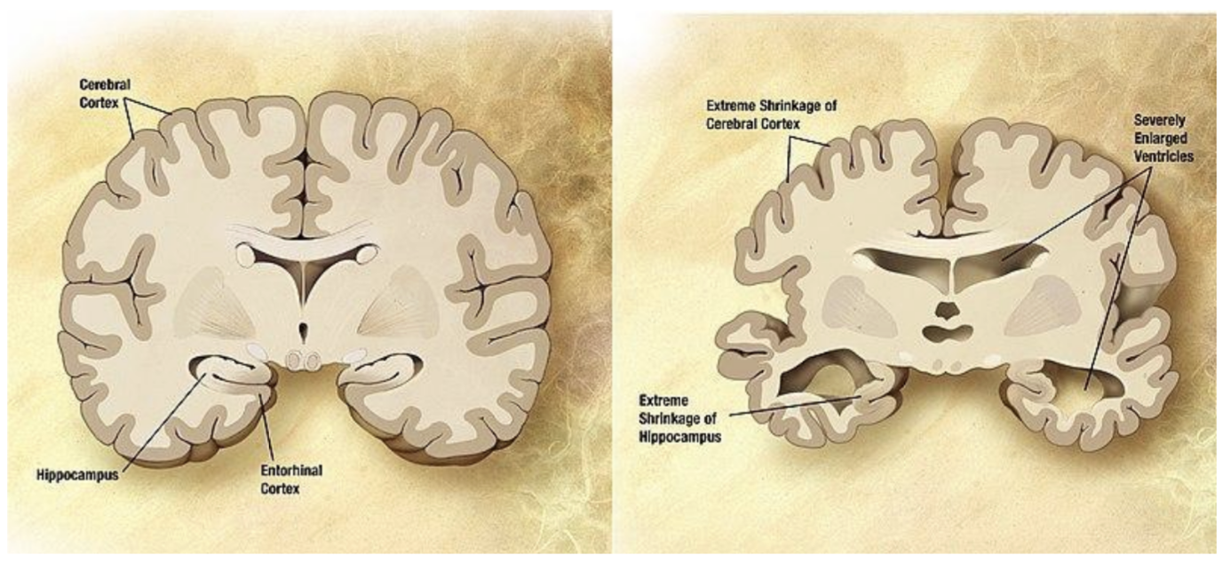
Image Source: Wikimedia Commons (2008). Alzheimer’s disease brain severe, Licensed under a creative commons licence. Available for free at: https://commons.wikimedia.org/wiki/File:Alzheimer%27s_disease_brain_severe.jpg (Accessed on: 18 April 2023).
Histopathological features of Alzheimer’s disease
| Senile plaques / Neuritic plaques | Neurofibrillary tangles |
| Develop before symptomatic cognitive impairment. | Develop at the same pace as cognitive decline. |
| Extracellular, beta amyloid globular structure along with surrounding degenerating neuronal processes. | Intracellular, formed due to the hyperphosphorylation of microtubule associated protein tau. |
| Poorly seen with H&E stain. Silver / Thioflavin S / Congo Red can be used. | Can be seen with H&E, easier to find with special stains such as silver or thioflavin S. |
| Origin of beta amyloid: Amyloid precursor protein (APP) is found on the cell membranes across the body, including the brain. It is cleaved by two enzymes: beta secretase and gamma secretase. |
Other neuropathological findings in Alzheimer’s disease
- Cerebral amyloid angiopathy (CAA) – Beta amyloid deposition in the small and medium sized arteries of the brain. This makes these vessels prone to rupturing and can lead to intracranial bleeding.
- Granulovacuolar degeneration (GVD).
- Hirano bodies – Rod like structures. It can also be found in other neurodegenerative diseases such as frontotemporal degeneration and CJD. These are eosinophilic in nature.
These can be found in healthy individuals as well. However, an increased prevalence is noted in patients with Alzheimer’s disease.
Neuropathological staging of Alzheimer’s disease
- 2012 NIA-AA Criterion – Divides Alzheimer’s into pre-clinical, mild cognitive impairment, and dementia caused by Alzheimer’s.
- Braak and Braak staging system – Uses only neuropathological components to assess severity.
- CERAD Criteria (The consortium to establish a registry for Alzheimer’s disease) – Clinical + neuropathological components.
(Don’t worry; the exams do not go into a lot of details about this, so just being familiar with the names is enough – for now!)
References
- Chan, G. (2021). Neuropathology: Part 1 – dementia. Trainees Online (TrON). Available online at: https://elearninghub.rcpsych.ac.uk/product?catalog=33_TRON_Neuropath_p1 [Accessed April 2023]
- Patient.info. (n.d.). What is Alzheimer’s Disease – Symptoms & Stages. Available online at: https://patient.info/doctor/alzheimers-disease. [Accessed March 2023]
Vascular dementia
Vascular dementia is the second most common dementia in adults over the age of 65. It is usually common that vascular dementia is ‘mixed’ with a coexisting pathology such as Alzheimer’s disease.
There are many types of vascular dementia, such as haemorrhagic dementia and multi-infarct dementia. Small vessels are involved in diseases such as CADASIL (Cerebral autosomal dominant arteriopathy with subcortical infarcts and leukoencephalopathy), which is the most common genetic cause for vascular dementia (autosomal dominant mutation of the notch 3 gene on chromosome 19).
There is a classical step-ladder type deterioration with vascular dementia.
NINDS-AIREN criteria for the clinical diagnosis of PROBABLE vascular dementia (VaD)
Presence of dementia – Cognitive decline from a higher level of functioning. This can be demonstrated as memory loss plus impairment in two or more different cognitive domains. This should be established by clinical examination and neuropsychological testing. Deficits should be severe enough to interfere with activities of daily living – not secondary effects of the cerebrovascular event alone.
Cerebrovascular disease – Defined by the presence of signs on neurological examination and / or by brain imaging.
A relationship between the above two disorders is inferred by:
- Onset of dementia within three months following a recognised stroke.
- An abrupt deterioration in cognitive functions.
- Fluctuating, stepwise progression of cognitive deficits.
Most vascular dementia is sporadic, but some cases display familial traits. Risk factors for vascular dementia include:
- History of stroke or transient ischaemic attack (TIA).
- Atrial fibrillation.
- Hypertension.
- Diabetes mellitus.
- Hyperlipidaemia.
- Smoking.
- Obesity.
- Coronary heart disease.
- Family history of stroke or cardiovascular disease.
Ischaemic changes include acute, sub-acute and chronic changes where the dead brain tissue is completely removed and replaced by scar tissue. This process is called gliosis.
References
- Chan, G. (2021). Neuropathology: Part 1 – dementia. Trainees Online (TrON). Available online at: https://elearninghub.rcpsych.ac.uk/product?catalog=33_TRON_Neuropath_p1 [Accessed April 2023]
- National Institute for Health and Care Excellence (2018). Overview | Dementia: Assessment, Management and Support for People Living with Dementia and Their Carers | Guidance | NICE. Available online at: https://www.nice.org.uk/guidance/ng97 [Accessed Feb 2023]
Frontotemporal dementia
Frontotemporal dementia (FTD) refers to a clinical syndrome of the following types:
- Semantic dementia (SD) – This could include a failure to recognise common objects. A patient might refer to a dog as a four-legged animal.
- Progressive non-fluent aphasia (PNFA) – Fragmented and broken speech, comprehension intact.
- Behavioural variant (bvFTD) – This is the most common type of FTD. This includes disinhibition, change in daily routines, and emotional dysregulation.
Since the frontal lobes are involved in emotions, behaviour, social intelligence and language, bvFTD would primarily affect these. The temporal lobes are involved with the comprehension of words. Hence, SD would primarily affect these.
So, what exactly is Pick’s disease?
FTD and Pick’s disease are often used interchangeably, but these are not exactly the same. Pick’s disease was described by Arnold Pick in 1892. Clinically, patients with Pick’s disease present similarly to patients with bvFTD. Pick’s disease is a tauopathy (excess tau proteins) and presents with behaviour changes following memory changes. In advanced stages, it often resembles Alzheimer’s disease closely.
An exam favourite is the classical ‘knife-blade’ atrophy of the frontal and temporal lobes, which shows the sharp delineation between atrophied and non-atrophied parts of the brain.
In histopathology, Pick’s disease has Pick bodies (cytoplasmic neuronal inclusions which have tau). These can be visualised well with a silver stain. This is also accompanied by neuronal loss, gliosis and microvacuolation.
Pick cells are DIFFERENT from Pick bodies – the former are swollen neurons with displaced nuclei.
In questions, terms like ‘ballooned neurons’ might be used.
References
- Alzheimer’s Society (2022). Alzheimer’s Society – United against dementia | Alzheimer’s Society. Available online at: https://www.alzheimers.org.uk/ [Accessed April 2023]
- Chan, G. (2021). Neuropathology: Part 1 – dementia. Trainees Online (TrON). Available online at: https://elearninghub.rcpsych.ac.uk/product?catalog=33_TRON_Neuropath_p1 [Accessed April 2023]
Lewy body dementia
These are named after the specific protein structures in this form of dementia called Lewy bodies.
Lewy body dementia (LBD) includes dementia with Lewy bodies and Parkinson’s disease dementia. If memory loss is present for more than a year before developing Parkinson’s disease, the diagnosis points towards dementia with Lewy bodies.
Symptoms of LBD include visual hallucinations, cognitive fluctuations, falls, sleep disturbance, movement disorders as seen with Parkinson’s disease, depression and an increased risk of neuroleptic side effects.
LBD primarily affects neurons of the substantia nigra in the midbrain. The special protein found in Lewy bodies is alpha-synuclein.
Antibodies to ubiquitin protease can be used to identify Lewy bodies. There is currently no correlation between the number of Lewy bodies and cognitive decline.
Lewy bodies can be present in the brainstem or cortex. In the brainstem, these are known as classical Lewy bodies and have a halo. These lewy bodies are the only histopathological lesions needed to make a pathological diagnosis of Lewy body dementia.
References
- Chan, G. (2021). Neuropathology: Part 1 – dementia. Trainees Online (TrON). Available online at: https://elearninghub.rcpsych.ac.uk/product?catalog=33_TRON_Neuropath_p1 [Accessed April 2023]
- Love, S. (2005). Neuropathological investigation of dementia: a guide for neurologists. Journal of Neurology, Neurosurgery, and Psychiatry 2005;76(Supplement 5 ):v8–‐‑v14.
Complete Guide to Clinical Genetics ST3 Application and Recruitment (2027)
Written by Medibuddy. Last updated: October 2026. Clinical Genetics is

Complete Guide to Clinical Radiology ST3 Application and Recruitment (2027)
Recruitment paused: NHS England is not running Clinical Radiology ST3+ recru

Complete Guide to Paediatric Surgery ST3 Application and Recruitment in United Kingdom (2026)
Written by Medibuddy. Last updated: October 2026. If you’re planning


Take your subscriptions with you
Our mobile app allows you to access your interview and exam question banks wherever you are.

