


 Close
Close
 11th April 2024
Sexual Dysfunction
Sexual dysfunction in males
Definition and prevalence
Male sexual dysfunction includes erectile dysfunction (ED), reduced libido and ejaculatory disorders. It is a common condition: 2 in 5 men in the UK experience ED and prevalence increases significantly between 40-70 years. As male arousal and sexual function are mediated by the interaction of hormonal, neurological, vascular and psychogenic factors, the causes of male sexual dysfunction are broad, and a comprehensive clinical history and examination are essential.
Erectile Dysfunction
Erectile dysfunction is a common problem defined as a persistent inability to attain and maintain an erection sufficient to permit satisfactory sexual performance. The incidence increases with age such that up to 50% of men will experience ED at ≥80 years. Its causes are broad (see Table 1) and can be best categorised into organic, psychogenic or mixed. Organic causes tend to present with a sub-acute or chronic onset, whereas psychogenic causes are often sudden or intermittent.
| Organic Causes | Psychogenic Causes | |
|---|---|---|
| Vascular | Cardiovascular disease | Anxiety |
| Diabetes mellitus | Depression | |
| Hypercholesterolaemia | Relationship dysfunction | |
| Hypertension | PTSD | |
| Peripheral vascular disease | Schizophrenia | |
| Smoking | Stress disorder | |
| Neurological | Multiple Sclerosis | |
| Parkinson’s disease | ||
| Radiotherapy | ||
| Spinal cord injury | ||
| Stroke | ||
| Anatomical | Epispadias / hypospadias | |
| Phimosis | ||
| Peyronie’s disease | ||
| Hormonal | Hyperprolactinaemia | |
| Hypogonadism | ||
| Thyroid disease | ||
| Drug Related | Antiarrhythmics (amiodarone, digoxin) | |
| Anti-depressants (SSRIs, lithium, TCAs) | ||
| Anti-hypertensives (β Blockers, CCBs, spironolactone) | ||
| Anti-psychotics (haloperidol) | ||
| Hormonal (goserelin, leuprorelin, finasteride) | ||
| Recreational (alcohol, cocaine, cannabis, opioids) | ||
Table 1. Causes of erectile dysfunction.
In addition to age, the factors associated with ED include cardiovascular disease, diabetes mellitus, dyslipidaemia, smoking and medication. As a result, the most common causes are vascular and drug-related, and ED is often a sign of peripheral or cardiovascular disease.
Investigations
The focus of investigations is on determining cardiac risk, associated metabolic syndromes and hormonal dysfunction. Blood tests should include: HbA1c, lipid profile, LFTs, TFTs, total testosterone and U&Es. Total testosterone should be performed as a fasting morning sample and repeated alongside FSH, LH, prolactin and sex hormone-binding globulin if found to be low. An ECG should be performed. If LUTS are present, a PSA should be considered.
Management
Lifestyle modification, including weight loss, smoking cessation, alcohol reduction and regular exercise, should be considered. Any associated cardiovascular (e.g. HTN) or metabolic (e.g. diabetes mellitus) conditions should be optimised. If a drug cause is suspected, a trial suspension can be considered (although most men benefit more from a PDE-5 inhibitor than drug suspension).
For all men not at high cardiac risk from sexual activity, a PDE-5 inhibitor such as sildenafil or tadalafil should be considered regardless of cause. PDE-5 inhibitors increase cyclic GMP in the corpus cavernosum through competitive inhibition of PDE-5, which results in nitric oxide-induced vasodilation. Onset is 30 – 60 minutes and the duration is 4 – 5 hours (up to 12 hours in some). Contraindications include a recent history of myocardial infarction, unstable angina or stroke, or retinitis pigmentosa. Profound hypotension can occur if co-administered with other vasodilators or anti-hypertensives. Common side effects include facial flushing, headaches, dizziness and short-term, blue-tinged vision. All men at high cardiac risk should be referred to Cardiology for assessment.
References
- National Institute for Health and Care Excellence (UK). Clinical Knowledge Summaries (CKS). Last revised May 2023. Available at https://cks.nice.org.uk/
- UpToDate. Epidemiology and Etiologies of Male Sexual Dysfunction. Last revised August 2022. Available at https://www.uptodate.com/contents/epidemiology-and-etiologies-of-male-sexual-dysfunction?search=sexual%20dysfunction%20male&source=search_result&selectedTitle=1~150&usage_type=default&display_rank=1#H1450745730.
- British National Formulary (BNF). Treatment Summaries: Erectile Dysfunction. Available at https://bnf.nice.org.uk/treatment-summaries/erectile-dysfunction/.
- Li, Jim Z et al. “Prevalence, Comorbidities, and Risk Factors of Erectile Dysfunction: Results from a Prospective Real-World Study in the United Kingdom.” International Journal of Clinical Practice vol. 2022 5229702. 20 Mar. 2022, doi:10.1155/2022/5229702.
- Hackett, Geoff et al. “British Society for Sexual Medicine Guidelines on the Management of Erectile Dysfunction in Men-2017.” The Journal of sexual medicine vol. 15,4 (2018): 430-457. doi:10.1016/j.jsxm.2018.01.023.
Premature Ejaculation
Premature ejaculation is defined as: (1) a brief ejaculatory latency (within 60 seconds of penetration), (2) inability to delay or control ejaculation, and (3) psychological distress. It can be split into lifelong or acquired forms, with acquired forms being strongly associated with psychogenic factors.
SSRIs are the main drug treatment. Dapoxetine, a short-acting SSRI, is licensed for ‘as required’ use in premature ejaculation. Other SSRIs (e.g., citalopram, fluoxetine, paroxetine, sertraline) are widely used as off-label treatments and are prescribed as regular medications which take 2 – 4 weeks to have effect. Topical anaesthetic preparations are also widely available.
References
- National Institute for Health and Care Excellence (UK). Clinical Knowledge Summaries (CKS). Last revised May 2023. Available at https://cks.nice.org.uk/.
- British National Formulary (BNF). Treatment Summaries: Premature Ejaculation. Available at https://bnf.nice.org.uk/treatment-summaries/premature-ejaculation/.
- Althof, Stanley E et al. “International Society for Sexual Medicine’s guidelines for the diagnosis and treatment of premature ejaculation.” The Journal of sexual medicine vol. 7,9 (2010): 2947-69. doi:10.1111/j.1743-6109.2010.01975.x.
Gynaecomastia
Background
Gynaecomastia is a benign proliferation of the glandular breast tissue, as opposed to pseudo-gynaecomastia, that is due to fat deposition. It occurs in 35% of men and is most common in men between 50 – 69, although it is also common in adolescence and infancy.
It is caused by an imbalance between circulating oestrogen and androgen. Androgens have an apoptotic and anti-proliferative effect on glandular breast tissues, whereas oestrogen promotes proliferation. Any increase in free circulating oestrogen or decrease in testosterone can therefore lead to gynaecomastia. The four main mechanisms through which this can occur are shown in the image below.
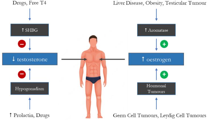
Image source: Authors’ Own Image
The cause is often multi-factorial and will vary depending on patient age. For instance, older males experience a decrease in testosterone production and an increase in adipose tissue, which increases the aromatisation of androgens into estrone (E1) and oestradiol (E2). Drugs are a common cause and act through suppression of androgen production, promotion of oestrogens or an increase in prolactin, resulting in hypogonadism.
The main causes are as follows:
| Drug Related | Anti-Androgenic Drugs | 5α Reductase (bicalutamide, finasteride, dutasteride) |
| Ketoconazole | ||
| Spironolactone | ||
| Antibiotics | Isoniazid | |
| Metronidazole | ||
| Cardiovascular Drugs | ACE-inhibitors | |
| Amiodarone | ||
| Calcium channel blockers | ||
| Statins | ||
| Chemotherapy Drugs | Alkylating agents | |
| Methotrexate | ||
| Vinca alkaloids | ||
| Gastrointestinal Drugs | Cimetidine | |
| Omeprazole | ||
| Psychoactive Drugs | Atypical antipsychotics (olanzapine, risperidone) | |
| Diazepam | ||
| Haloperidol | ||
| Tricyclic antidepressants | ||
| Drugs of Abuse | Alcohol | |
| Marijuana | ||
| Other | Domperidone, metoclopramide | |
| HAART | ||
| Phenytoin | ||
| Gonadal Failure | Hypogonadism | |
| Hyperprolactinaemia | ||
| Hormone Secreting Tumours | Adrenocortical malignancy | |
| Testicular malignancy | ||
| Other | Cirrhosis | |
| Chronic kidney disease | ||
| Hyperthyroidism | ||
| Obesity | ||
Table 2. Causes of gynaecomastia
Investigations
Initial blood tests should include morning testosterone, LFTs, TFTs and U&Es. If testosterone is low, then it should be repeated alongside FSH, prolactin, oestradiol and SHBG. If the history or examination is suggestive of testicular malignancy, then β-hCG, AFP and LDH should be performed alongside testicular ultrasound.
Management
All men with pathological gynaecomastia should be referred for specialist advice. Adolescents should not be treated unless symptoms have been present for at least 2 years, as in most cases, gynaecomastia is transient in this age group. Men whose gynaecomastia is likely secondary to drugs should be offered alternative treatment.
Medical management has a high success rate but is ineffective once breast fibrosis is established. Danazol is a synthetic steroid with anti-gonadotrophic and anti-estrogenic effects that is licensed to treat gynaecomastia. However, it is also associated with weight gain, which can exacerbate symptoms. Common off-licence medications include tamoxifen (a selective oestrogen receptor modulator) and aromatase inhibitors (e.g. letrozole, anastrozole). Tamoxifen should be avoided in patients with a history of venous thromboembolism.
Surgical intervention should be considered for men with fibrotic gynaecomastia.
References
- National Institute for Health and Care Excellence (UK). Clinical Knowledge Summaries (CKS). Last revised May 2023. Available at https://cks.nice.org.uk/.
- Thiruchelvam P, Walker J N, Rose K, Lewis J, Al-Mufti R. Gynaecomastia BMJ 2016; 354 :i4833 doi:10.1136/bmj.i4833.
- UpToDate. Clinical features, diagnosis and evaluation of gynaecomastia in adults. Last revised April 2023. Available at https://www.uptodate.com/contents/clinical-features-diagnosis-and-evaluation-of-gynecomastia-in-adults?search=gynecomastia&source=search_result&selectedTitle=1~150&usage_type=default&display_rank=1
Sexual dysfunction in females
Definition and prevalence
Female sexual dysfunction includes a spectrum of disorders that are often multi-factorial in nature: sexual interest and arousal disorder (SIAD), female orgasmic disorder (FOD) and genito-pelvic penetration pain disorder (GPPPD), which includes dyspareunia and vaginismus. The exact prevalence of female sexual dysfunction is uncertain, as most studies have historically focused on male sexual dysfunction, but it is estimated to be between 11% overall and 41% in premenopausal women. There are 3 criteria for diagnosing a sexual disorder: (1) symptoms present for at least 6 months; (2) symptoms are experienced in ≥75% of sexual encounters; (3) symptoms have caused significant distress.
The aetiology of sexual dysfunction is often multifactorial and involves psychological, contextual, pharmacological and physiological factors. For instance, SIAD is strongly linked to mood disorders, such as depression and anxiety, in addition to PTSD and previous experience of abuse. GPPPD can be predominantly psychological in origin (as in vaginismus) but can also be due to surgical, medical and pharmacological causes (see Table 3).
| Drug-Related | SSRIs | Aromatase inhibitors (letrozole, anastrozole) |
| Anti-hypertensives (β blockers, spironolactone, thiazides) | Lithium | |
| Anti-psychotics | Opioids | |
| Anti-epileptics | ||
| Medical | Diabetes mellitus | Perimenopause/menopause |
| Hyperthyroidism | Pelvic inflammatory disorder (PID) | |
| Hyperprolactinaemia | STI | |
| IBS/IBD | Thyroid disorders | |
| Interstitial cystitis | UTI | |
| Neurological disease (MS, Parkinson’s) | ||
| Surgical | Adenomyosis | Interstitial cystitis |
| Endometriosis | Malignancy (endometrial, cervical) | |
| FGM | Ovarian cyst | |
| Fibroids | Previous pelvic surgery | |
Table 3. Non-psychological causes of female sexual dysfunction.
References
- UpToDate. Overview of sexual dysfunction in females: Epidemiology, risk factors and evaluation. Last revised May 2022. Available at https://www.uptodate.com/contents/overview-of-sexual-dysfunction-in-females-epidemiology-risk-factors-and-evaluation?search=sexual%20dysfunction%20in%20women&source=search_result&selectedTitle=2~150&usage_type=default&display_rank=2#H12.
- BMJ Best Practice. Sexual dysfunction in women. Last revised December 2022. Available at https://bestpractice.bmj.com/topics/en-gb/352.
- Burri, A., & Spector, T. (2011). Recent and lifelong sexual dysfunction in a female UK population sample: prevalence and risk factors. The journal of sexual medicine, 8(9), 2420–2430. https://doi.org/10.1111/j.1743-6109.2011.02341.x.
Fibroids
Background
Fibroids are benign tumours that can develop in any part of the myometrium. They typically develop in women of reproductive age as their growth is stimulated by oestrogen and progesterone. As a result, the risk factors are those associated with increased oestrogen and progesterone exposure: (1) increased age (until menopause); (2) early menarche; (3) nulliparity; (4) obesity. Fibroids are more common in women of black and Asian descent and those with a family history in a first-degree relative.
Clinical evaluation
Most fibroids are asymptomatic and are discovered as incidental findings. Symptomatic fibroids may present with the following: (1) menorrhagia; (2) dysmenorrhoea; (3) dyspareunia; (4) abdominal bloating or discomfort; (5) lower urinary tract symptoms (LUTS); (6) recurrent UTIs; (7) constipation or painful defaecation; (8) subfertility or recurrent miscarriage. Patients may present with features of the complications of fibroids, such as iron deficiency anaemia (fatigue, exertional breathlessness) or, more rarely, hydronephrosis (dysuria, flank pain, oligo-anuria).
Investigations
If iron deficiency anaemia is suspected, then FBCs and iron studies should be ordered. Transabdominal and transvaginal ultrasound are used to confirm the presence of fibroids.
Management
All patients with features suggestive of gynaecological cancer (see Table 4) should be referred for a two-week wait assessment. Referral for specialist advice should be considered if there is heavy menstrual bleeding, fertility issues, fibroids measuring ≥3cm or rapid growth in post-menopausal women.
| Cervical Cancer | Appearance of cervix consistent with cervical cancer |
| Endometrial Cancer | Post-menopausal bleeding in women ≥55 |
| Ovarian Cancer | Any abdominal or pelvic mass which is not obviously uterine fibroids |
| Ascites and/or a pelvic or abdominal mass identified by physical examination which is not obviously uterine fibroids | |
| Vaginal Cancer | Unexplained vulval bleeding |
| Unexplained vulval lump or ulceration | |
| Unexplained and palpable vaginal mass | |
Table 4. Criteria for two-week wait referrals for suspected gynaecological malignancy. Source: NICE.
Medical management options include the treatment of menorrhagia with tranexamic acid, NSAIDs, the combined oral contraceptive pill or oral progestogen. Tranexamic acid is an anti-fibrinolytic that inhibits plasminogen conversion and thus reducing bleeding. NSAIDs reduce uterine blood loss via the inhibition of prostaglandin synthesis. Both the copper coil (Cu-IUD) and Mirena (LNG-IUS) are suitable for use in women with fibroids without distortion of the uterine cavity and often result in a decrease in symptoms and fibroid size. Hormonal contraception reduces the size of fibroids via the negative feedback loop within the HPG axis, in which increased oestrogen and progesterone suppress the production of FSH and LH that, in turn, inhibits ovulation (see image below).
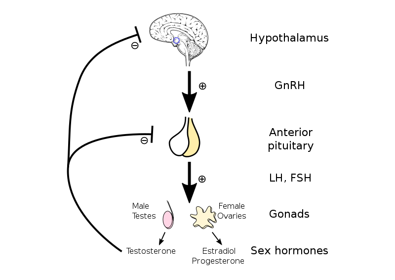
Image source: Wikimedia Commons (2019). Hypothalamic–pituitary–gonadal axis, Licensed under a Creative Commons licence. Available for free at: https://commons.wikimedia.org/wiki/File:Hypothalamic%E2%80%93pituitary%E2%80%93gonadal_axis.svg (Accessed: 07 February 2024).
Gonadotropin-releasing hormone (GnRH) agonists such as leuprorelin and goserelin may be used to shrink uterine fibroid size prior to surgical intervention. These function by initially increasing the production of gonadotrophins, which results in receptor desensitisation, leading to a hypogonadal state that simulates menopause.
References
- National Institute for Health and Care Excellence (UK). Clinical Knowledge Summaries (CKS). Last revised May 2023. Available at https://cks.nice.org.uk/.
- UpToDate. Uterine fibroids (leiomyomas): Epidemiology, clinical features, diagnosis and natural history. Last revised on 14 June 2023. Available at https://www.uptodate.com/contents/uterine-fibroids-leiomyomas-epidemiology-clinical-features-diagnosis-and-natural-history?search=fibroid&source=search_result&selectedTitle=2~150&usage_type=default&display_rank=2.
- The Faculty of Sexual and Reproductive Healthcare. UK Medical Eligibility Criteria for Contraceptive Use (UKMEC). Last revised 2019. Available at https://www.fsrh.org/documents/ukmec-2016/.
- Kessel, B., Liu, J., Mortola, J., Berga, S., & Yen, S. S. (1988). Treatment of uterine fibroids with agonist analogs of gonadotropin-releasing hormone. Fertility and sterility, 49(3), 538–541.
Female genital mutilation (FGM)
Background
FGM describes any procedure involving the partial or total removal of the external genitalia or any other injury to the female genital organs for non-medical reasons. It is estimated that 65,000 girls aged 13 and under in the UK are at risk of FGM. It is most commonly practised among African (Egypt, Nigeria, Kenya, Sierra Leone, Somalia, Sudan) and some non-African (Afghanistan, Indonesia, Kurdistan, Malaysia, Turkey, Thailand) communities.
Clinical evaluation
Symptoms that suggest FGM is present are: (1) difficulties walking, standing or sitting; (2) urinary retention and/or incontinence; (3) menstrual problems; (4) vaginal soreness or recurrent infections; (5) depression or withdrawn behaviour. Where FGM is suspected, women should be offered STI screening, including HIV, Syphilis, hepatitis B and C. All women with FGM should be referred to FGM services (where available) or secondary care.
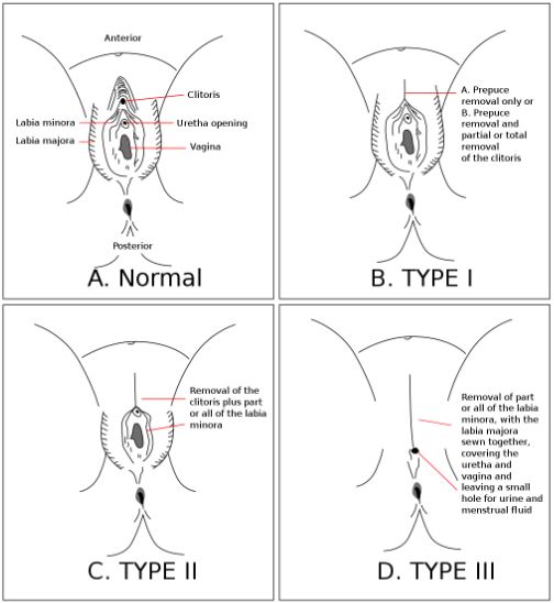
Image source: Wikimedia Commons (2020). FGC Types, Licensed under a Creative Commons licence. Available for free at: https://commons.wikimedia.org/wiki/File:FGC_Types.svg (Accessed: 07 February 2024).
Legal duties
FGM has been a criminal offence in the UK since 1985. It is illegal to arrange, assist in arranging or fail to protect a girl from the risk of FGM. Since 2014, it has been mandatory for all healthcare professionals to record the identification of FGM within a patient’s clinical record and to submit these details to the DHSC. While there is no requirement to ask every female whether they have FGM, healthcare staff are expected to use professional judgement to decide whether there are risk factors for each patient.
All vulnerable adults or children under 18 who have signs of FGM, or in whom there is good reason to suspect they are at risk of FGM, must be referred to the police and child social services within 1 month. There is no requirement to refer adult women to social services or the police.
References
- Department of Health and Social Care. Multi-agency statutory guidance on female genital mutilation (1 April 2016). Last revised on 30 July 2020. Available at https://www.gov.uk/government/publications/multi-agency-statutory-guidance-on-female-genital-mutilation.
- Royal College of Obstetricians and Gynaecologists. Female Genital Mutilation and its Management (Green-top Guidelines No. 53). July 2015. Available at https://www.rcog.org.uk/media/au0jn5of/gtg-53-fgm.pdf.
Complete Guide to Clinical Genetics ST3 Application and Recruitment (2027)
Written by Medibuddy. Last updated: October 2026. Clinical Genetics is

Complete Guide to Clinical Radiology ST3 Application and Recruitment (2027)
Recruitment paused: NHS England is not running Clinical Radiology ST3+ recru

Complete Guide to Paediatric Surgery ST3 Application and Recruitment in United Kingdom (2026)
Written by Medibuddy. Last updated: October 2026. If you’re planning


Take your subscriptions with you
Our mobile app allows you to access your interview and exam question banks wherever you are.

