


 Close
Close
 26th June 2023
Spinal cord syndromes
Complete Cord Transection

Distribution of motor and sensory loss in complete lower thoracic spinal cord transection.
Image Source: Dr D Chang – own work
In a complete cord transection, there is a loss of motor function (corticospinal), pain and temperature sense (spinothalamic), and touch and proprioception sense (dorsal column) below the lesion, which can result in paraplegia or tetraplegia with loss of all sensory input depending on the level of the lesion. The affected limbs will initially be flaccid and areflexic due to spinal shock before progressing to hypertonic and hyper-reflexive clinical findings with bilateral upgoing plantar reflexes (Babinski’s sign).
Autonomic dysfunction is also a feature, with disruption to urinary and rectal sphincter functions mediated by sympathetic/parasympathetic pathways, which can manifest as anhidrosis, impaired temperature control and loss of the cremasteric reflex (stroking of the medial thigh causing the cremaster muscle to contract and pull up the ipsilateral testicle toward the inguinal canal.)
Its aetiology is primarily due to traumatic and inflammatory causes. Inflammatory causes include multiple sclerosis and postinfectious inflammation from viral, bacterial, and fungal causes, and it can be a very rare complication of vaccination.
It can also result from trauma, such as a traumatic spinal injury or herniated intervertebral disc, and other mechanical causes, such as tumour and metastatic disease.
Prognosis of recovery in traumatic cases is poor with a less than 5 percent chance of recovery of function; recovery in nontraumatic cases is dependent on aetiology.
Brown-Séquard Syndrome
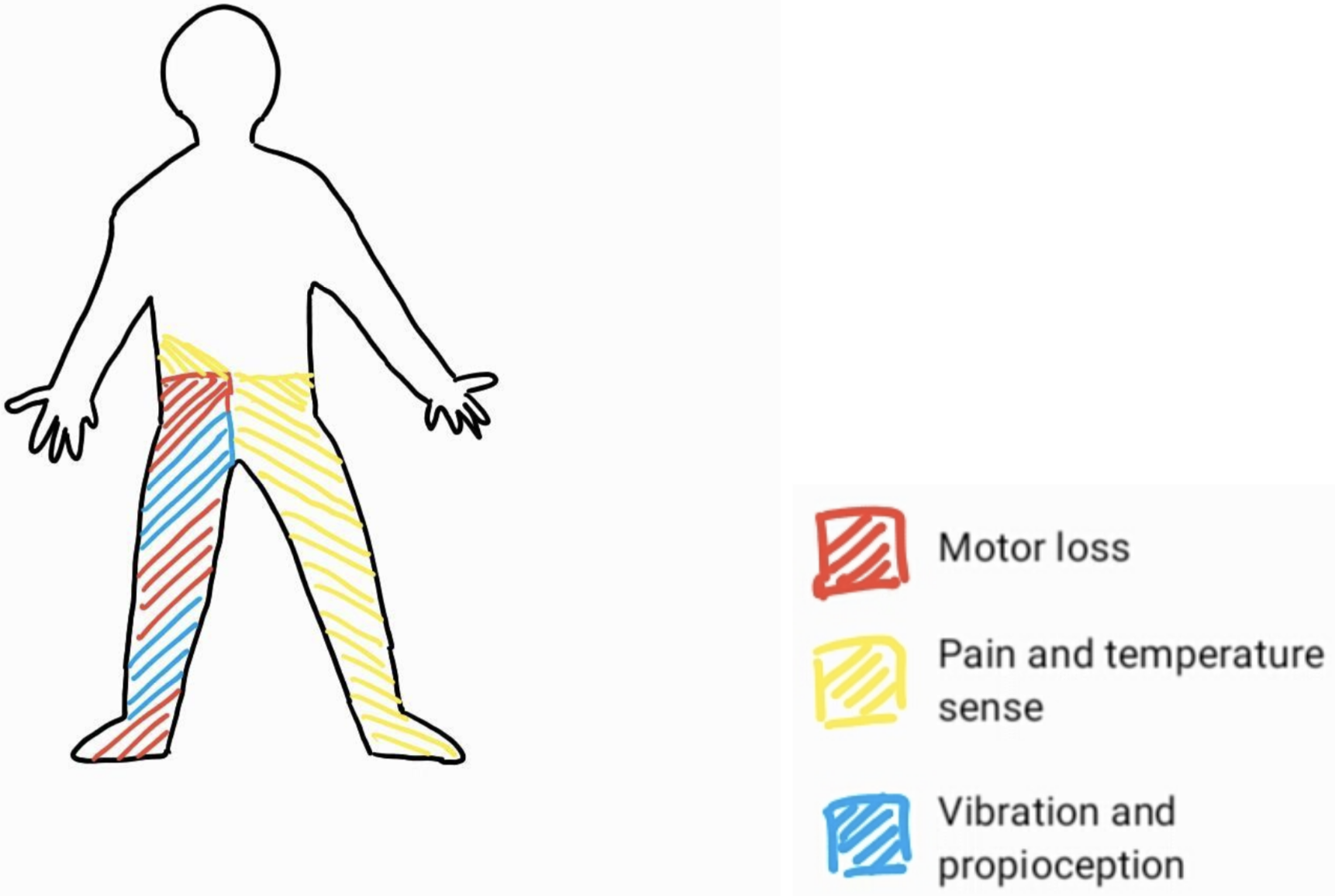
Distribution of motor and sensory loss in right hemisection of the lower thoracic spinal cord.
Image Source: Dr D Chang – own work
Brown-Séquard syndrome describes a pattern of sensorimotor loss resulting from damage to one half of the spinal cord (i.e. hemisection of the cord) causing:
- Ipsilateral loss of vibration sense and proprioception, with hemiparesis.
- Contralateral loss of pain and temperature sense, with a clinical sensory level usually 1 to 2 levels below the actual lesion.
- Ipsilateral loss of all sensory modalities at the level of the lesion, with radicular pain.
- Ipsilateral flaccid paralysis at the level of the lesion.
Brown-Séquard syndrome is usually caused by a traumatic spinal injury or intramedullary tumours confined to one side of the spinal cord, but an important cause of Brown-Séquard syndrome is transverse myelitis (see below).
This syndrome has the most favourable prognosis among the incomplete spinal cord syndromes, with 75% of patients able to ambulate independently at discharge from rehabilitation (75%) and nearly 70% perform functional skills and activities of daily living independently and favourable bladder and bowel function profile with continent bladder and bowel function achieved in 89% and 82%, respectively.
Central Cord Syndrome

Distribution of motor and sensory loss in central cord syndrome from a large lesion affecting the corticospinal tracts, spinothalamic tracts, and dorsal columns at the level of the cervical spine.
Image Source: Dr D Chang – own work
Central cord syndrome is the most common incomplete spinal cord syndrome and has a varied presentation depending on how large the lesion is, which determines how much cord is involved. It is insidious in onset, affecting first the anterior and lateral spinothalamic tracts, causing loss of pain and temperature with preservation of position, vibration, and touch sensation. Lesions causing central pattern spinal cord syndrome typically arise at the level of the cervical spinal cord. Sensory loss here presents in a vest-like pattern over the shoulders with a spectrum of range and severity with size of lesion. Sacral sensation is usually spared.
Larger lesions can progress posteriorly to include the dorsal columns, affecting vibration and proprioception below the lesion, anteriorly to the anterior horns, and laterally involving the corticospinal tracts, causing a flaccid paralysis with fasciculations, with eventual atrophy at the level of the lesion and spastic paralysis of muscles below the lesion.
The aetiology of a central pattern syndrome is most commonly from a traumatic hyperextension injury involving the cervical cord, which specifically injures the central part of the cord, typically the central corticospinal tracts (as opposed to fractures/trauma causing other complete or incomplete spinal cord syndromes). This can result in quadriplegia with some recovery of lower limb function and a disproportionately greater upper limb weakness, worse distally than proximally. This is more common in people with cervical osteoarthritis and cervical spondylosis, where an existing stenosis, e.g. from osteophytes, increases the vulnerability of the spinal cord to injury. Bladder dysfunction, e.g. urinary retention, priapism and autonomic dysreflexia may occur.
In traumatic central cord syndrome, the prognosis is generally favourable, with patients recovering substantial neurological function after the injury with 90 percent return of motor function by three years, progressing in an ascending order with return of lower limb function, then bladder control and eventually proximal upper limbs and hands. Patients undergoing surgical intervention are more likely to attain pre-injury mobility status.
Atraumatic causes are much rarer and include neoplastic causes such as intramedullary spinal cord tumours and syringomyelia.
Syringomyelia

Distribution of pain and temperature loss in syringomyelia.
Image Source: Dt D Chang – own work
Syringomyelia is the development of a CSF filled syrinx within the spinal cord parenchyma or central canal that can enlarge and compress surrounding tracts, typically at the C8/T1 level. It is typically associated with type 1 Arnold-Chiari malformations in which there is downward displacement of the cerebellar tonsils beneath the foramen magnum into the cervical spinal canal, blocking the normal CSF flow between the spinal canal and intracranial space. Syringomyelia can also be associated with a tethered cord (in which the caudal spinal cord can become fixed to surrounding inelastic tissue) and with spina bifida, tumours, meningitis and arachnoiditis.
Syringomyelia typically presents with bilateral pain and temperature insensitivity in a cape-like distribution over the upper limbs due to the compression of the spinothalamic tract decussation in the anterior white commissure, with preserved touch and vibration sense below the level of the syrinx. Symptoms can be static for years but may worsen in association with Valsalva manoeuvres, e.g. coughing or sneezing/heavy lifting/straining, which can cause enlargement and extension of the syrinx.
Enlargement of the syrinx causes the progression of the central cord syndrome, compressing anterior horn cells and corticospinal tracts, leading to a variable presentation of syringomyelia depending on the size of the syrinx and which structures are being compressed. This can include spasticity or paralysis of the legs, ataxia, or fasciculations.
If the syrinx is higher in the cord, extending into the medulla of the brainstem, this is termed syringobulbia, which can result in cerebellar and lower motor cranial nerve nuclei signs.
Investigation is with MRI and dynamic CSF flow MRI studies, with CT myelograms as an alternative in the event of MRI contraindications.
Treatment is typically with surgical intervention to improve CSF flow and is dependent on the cause, e.g. a Chiari malformation indicating a craniocervical decompression if associated with type 1 Arnold-Chiari malformation, or with decompressive laminectomy, syringosubarachnoid or syringoperitoneal shunts.
Posterior Column Syndrome

Distribution of vibration and proprioception loss in posterior column syndrome of the cervical spinal cord.
Image Source: Dr D Chang – own work
Posterior column syndrome is the most uncommon incomplete spinal cord syndrome, manifesting as a loss of proprioception and vibratory sense, and two-point discrimination distal to the lesion, deep tendon areflexia and a sensory ataxia (due to loss of joint position sense). Romberg sign is positive, and a stomping gait is present which can be exacerbated by removing visual cues, e.g. by closing eyes. There is hypotonia in limbs distal to the lesion, but power is usually preserved. If the lesion is at the level of the cervical cord, it can be associated with Lhermitte’s sign, in which neck flexion elicits neuropathic pain and paraesthesia down the arms.
The aetiology or posterior cord syndrome is classically due to neurosyphilis with tabes dorsalis, but can happen due to thrombosis or occlusion of the posterior spinal arteries, mechanical causes such as localised compression from epidural tumours, haematomas or abscesses, and inflammatory causes such as multiple sclerosis. Friedreich’s ataxia (see the section below) can also manifest primarily as a posterior cord syndrome before progressing to involve corticospinal tracts.
In the case of tabes dorsalis as a late complication of neurosyphilis, posterior column degeneration is also accompanied by dorsal root ganglion demyelination. Tabes dorsalis progressively develops over years, manifesting with the posterior cord syndrome as above, accompanied with lancinating pain and dysesthesias and can progress to cause bladder dysfunction, Argyll Robertson pupils (see Cranial Nerve Disorders section) and can cause a Charcot arthropathy. Treatment is with intramuscular benzylpenicillin, noting that patients undergoing treatment may experience a Jarisch Herxheimer reaction where the death of the Treponema pallidum spirochetes release endotoxins that provoke pyrexia, tachycardia, rigors, and rashes 6 to 48 hours post initiation of therapy.
Posterolateral Column Syndrome
Posterolateral column syndrome, also known as ‘Subacute Combined Degeneration of the spinal cord’ (SACD), is similar to a posterior column syndrome, but also involves the lateral corticospinal tracts. This is a slow and insidious spinal cord syndrome characterised by ascending bilateral limb paraesthesia, manifesting as loss of position and vibratory sense, progressing to weakness with ataxia, and eventually a bilateral spastic paresis. Pain and temperature sense is unaffected. Clinical findings may show a sensory ataxia with positive Romberg’s sign and bilateral spastic paresis with a positive Babinski sign.
It is usually caused by metabolic aetiology, most commonly a prolonged vitamin B12 deficiency. This is why a patient with folate deficiency should always have B12 levels checked, as symptoms of B12 deficiency can be exacerbated and masked if only the folate deficiency is treated. Subacute Combined Degeneration of the spinal cord can also be caused by vitamin E deficiency and copper deficiency, causing loss of myelin initially at the dorsal columns (governing vibration, coarse touch, and proprioception) and then at the lateral columns, including spinocerebellar and corticospinal tracts, but sparing the spinothalamic tracts.
Diseases which exacerbate these nutritional deficiencies, e.g. pernicious anaemia causing vitamin B12 deficiency, cystic fibrosis or abetalipoproteinemia causing loss of absorption of vitamin E, or Menkes disease, an X-linked recessive mutation in copper transport proteins, can all cause SACD. Of note, nitrous oxide administration (e.g. as anaesthesia, analgesia, or as a recreational drug) can induce a functional B12 deficiency and precipitate SACD in already B12 deficient individuals, by inactivating vitamin B12. This is assessed by checking methylmalonic acid (MMA) and homocysteine levels.
Treatment of SACD is with intramuscular hydroxocobalamin injections to replace vitamin B12 levels.
Anterior Cord Syndrome

Distribution of motor and sensory loss in anterior cord syndrome of the cervical spinal cord.
Image Source: Dr D Chang – own work
Anterior cord syndrome is an incomplete spinal cord syndrome affecting the anterior two-thirds of the spinal cord. In general, an anterior cord syndrome manifests as bilateral motor paralysis below the level of the lesion, with associated bilateral loss of pain and temperature sense. Lateral horn involvement can cause an autonomic dysfunction, including anhidrosis, hypotension, neurogenic bowel, bladder, and sexual dysfunction.
There are two primary ‘sub-syndromes’ depending on whether the anterior horn cells are affected (anterior horn cell syndrome) or whether the vascular supply to the anterior cord is the aetiology of the syndrome (anterior spinal artery syndrome).
In anterior horn cell syndrome, there is involvement of the anterior grey matter horns of the spinal cord, causing ipsilateral or bilateral flaccid motor paralysis with atrophy and fasciculations. The aetiology is varied and includes:
- Infections: Poliomyelitis, West Nile virus
- Iatrogenic: Post-radiation myelitis
- Metabolic: Hexosaminidase A deficiency (aka lysosomal storage disorder), GM2 gangliosidosis (aka Tay Sachs disease)
- Hereditary: Spinal muscular atrophies
Anterior spinal artery syndrome is typically vascular in aetiology, with disruption of the anterior spinal artery or the great artery of Adamkiewicz from a thrombus, ruptured aortic aneurysm, aortic dissection, or vasculitis. Disruption of vascular supply, e.g. in severe hypotension or a cocaine associated vasospasm, can cause a similar presentation. Onset is abrupt with deficits occurring minutes to hours post insult. Clinical features may include radicular back or neck pain and acute flaccid paralysis with loss of pain and temperature and autonomic dysfunction. Symptoms may progress to bilateral spastic para/quadriparesis over days to weeks. Touch, proprioception, and vibration sense are preserved due to the sparing of the dorsal column medial lemniscus tracts.
Treatment involves management of the underlying cause, e.g. fluid/vasopressors for hypotension, as well as supportive therapies, including physiotherapy and occupational therapy, social and psychological support along with pressure area care, bladder catheterisation, venous thromboembolism prophylaxis and stress ulcer protection. Prognosis is poor and many patients do not recover motor, sensory or autonomic function.
Complete Guide to Clinical Genetics ST3 Application and Recruitment (2027)
Written by Medibuddy. Last updated: October 2026. Clinical Genetics is

Complete Guide to Clinical Radiology ST3 Application and Recruitment (2027)
Recruitment paused: NHS England is not running Clinical Radiology ST3+ recru

Complete Guide to Paediatric Surgery ST3 Application and Recruitment in United Kingdom (2026)
Written by Medibuddy. Last updated: October 2026. If you’re planning


Take your subscriptions with you
Our mobile app allows you to access your interview and exam question banks wherever you are.

